
Musculoskeletal Pearls
- Intra-articular fractures with 25% or greater involvement of the joint surface should be referred to orthopedics for surgical treatment.
- Musculoskeletal Red Flags.
| Features | |
| SCFE (Slipped Capital Femoral Epiphysis) | -Occurs more commonly during the adolescent growth spurt (11-13 years of age for girls, 13-15 years for boys) when the femoral head is displaced posteriorly through the growth plate. -Risk factors: Femoral retroversion or steeper inclination of the proximal femoral physis; being overweight/obese; being African American/Islander; being male (more common in males than females), and being physically active Pt may present with a limp and pain in the groin or anterior thigh but also may present with referred pain to the knee. Others present with a painless limb or vague pain.Once the diagnosis of SCFE is made, the patient should not bear weight and should be referred promptly for surgery to prevent complications. On PE, limited internal rotation of the flexed hip is pathognomonic for SCFE. Specific to SCFE is even greater limitation of internal rotation when the hip is flexed to 90 degrees. No other pediatric condition has this typical finding, which makes the maneuver very useful in children with LE pain. Orthopedic consultation is advised if SCFE is suspected. |
| Avascular or aseptic necrosis of the femoral head (Legg-Calve-Perthes disease). | -Commonly occurs in boys 4-8 years of age. -In addition to hip (or knee pain), limping is a prominent feature. |
| Meralgia Paresthetica | -Upper thigh numbness in an adolescent female is a classic symptom of Meralgia Paresthetica. It is caused by impingement of the lateral femoral cutaneous nerve in the origin, 2/2 to obesity or wearing clothes too tight in the waist or groin. |
| Developmental dysplasia of the hip | |
| Polymyositis/Dermatomyositis | Proximal muscle weakness, elevation of CK and aldolase. |
| Amyotrophic Lateral Sclerosis | |
| Myasthenia gravis | |
| Duchenne’s muscular dystrophy | |
| A flexor digitorum profundus avulsion fracture”Jersey Finger”. | Results from forced hyperextension fo a flexed DIP joint. On PE, the patient is unable to flex the finger at the DIP joint. Radiographs will show a bony fragment at the volar surface of the proximal distal phalanx.
Refer patient to a hand surgeon as soon as the diagnosis is made b/c the risk of tendon retraction is high. |
| Paget’s disease of bone. | -Bone pain is continuous. Unlike the pain of OA, bone pain of Paget’s disease usually increases with rest, when the limbs are warmed, and at night. Bowing may occur in Tibia. Alkaline Phosphatase is elevated. |
| Seronegative Spondyloarthritis (spondyloarthropathy) | -Is a family of inflammatory rheumatic diseases that cause arthritis. The most common is ankylosing spondylitis, which affects mainly the spine. |
| Boxer’s fracture | |
| Greenstick fracture | |
| Cole’s fracture | |
| Salter-Harris type II fracture | See Salter-harris classification here |
| Spiral fracture | |
| FABER test | |
| FADIR test | |
| Malignant bone pain | Night pain is common |
| Sacroilliitis | Positive FABER test. |
| Transient synoviitis | |
| Muscle strain | |
| Benign nocturnal hip pains of childhood (growing pains) | -Occur in as many as 1/3 of children, most common b/n 4 and 6 years of age. |
| Scaphoid Fracture | Wrist extension is decreased; tenderness over the dorsum of the wrist, particularly just distal and dorsal to the radial styloid. A dorsiflexion injury will typically cause a scaphoid fracture in a young adult, resulting in tenderness to palpation over the anatomic snuffbox. A plain posterior-anterior wrist radiograph is often normal. However, a special view with the wrist prone in ulnar deviation elongates the scaphoid, often showing subtle fractures. |
| Hook of the Hamate fractures | Cause tenderness at the proximal hypothenar area 1cm distal to the flexion crease of the wrist |
| Scapholunate dislocation | Can be identified with a “clenched-fist” view and supinated view with the wrist in ulnar deviation. |
| Lateral Epicondylitis (tennis elbow) – also called, lateral epicondylalgia to reflect the non-inflammatory nature of the condition. | This is an overuse tendinopathy of the common extensor tendon origin of the lateral elbow.
Modifying work routine (offloading the involved tendons) is the best way to get long-term better outcomes. |
| Vitamin D supplementation | |
| Iron supplementation in completely breastfed children. | |
| Eccentric strengthening excercises | =first line treatment for chronic Achilles tendinopathy. |
| Reducing a patella that is laterally dislocated | Apply a medially directed pressure on the patella while extending the leg. Have the patient sit or lie with the leg in a flexed position and then apply gentle medial pressure to the patella until the most lateral edge is over the femoral condyle. The leg should then be gently extended and the knee brought into full extension. This should cause the patella to slip back into place, and the knee should then be immobilized. |
| Acute low back pain | -Muscle relaxants like cyclobenzaprine (Flexeril) are beneficial for the relief of acute low back pain for the first 7-14 days after the onset of symptoms. -Topic NSAIDs like Voltaren gel also work well. |
| The major indication for knee replacement is | Severe joint pain. Other things can be considered, but severe joint pain is the main one. |
| Side effects of Bisphosphonates used to tx osteoporosis | |
| Calcium supplementation | -Calcium carbonate is the least expensive and most commonly used supplement, but it is constipating and stomach acid improves its absorption. -Calcium citrate is less dependent on stomach acidity for absorption and is good for people who are taking a PPI or H2 receptor blocker chronically. It may also be taken without regard to food or meals. |
| The most common source of chest pain in children is: | Musculoskeletal (50-60%) Psychogenic (10-30%) Respiratory (3-12%) Cardiac conditions (0-5%) |
| Plantar fasciitis | Diagnose with history and PE alone. Heel pain that is worse with the first steps in the morning is very suggestive of plantar fascitis. Plantar fasciitis is characterized by pain that is worse with the first few steps in the morning or after a prolonged rest. The pain is typically in the medial heel. 50% of people with plantar fasciitis have heel spurs on radiographs, however, this finding is not causative or diagnostic. The diagnosis is made clinically.
**Plantar fasciitis causes pain on the plantar surface of the heel rather than posteriorly, as in the case of calcaneal apophysitis (Sever’s disease). |
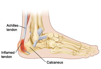
| Achilles Tendinopathy | Achilles tendinopathy causes tenderness to palpation of the Achilles tendon. |
| Tarsal Tunnel Syndrome | Is related to compression of the posterior tibial nerve causes neuropathic pain and numbness in the posteromedial ankle and heel. |
| Patellofemoral Pain Syndrome (PFPS) | Causes anterior knee pain that is worse with running downhill. PE is often normal, although there may be apprehension when the knee is extended with pressure over the patella and the patella will sometimes track laterally.
PFPS can be treated with exercises to strengthen the quadriceps and hips, and by using a knee sleeve with a doughnut-type cushion that the patella fits into. Management of PFPS: Am Fam Physician. 2007;75(2):194-202 |
| Pes anserine bursitis | |
| Stretching helps with: | Hamstring strain, chronic neck pain, Osteoarthritis, rehabilitation post knee replacement |
| Stretching has no benefits for | Patients with joint contracture |
| Lachman Test | A positive Lachman test indicates that the anterior cruciate ligament may be torn. |
| The McMurray Test | The McMurray and Thesally assessments test for meniscal tears. |
| Thessaly Test | |
| The Balottment test | Is for detecting intra-articular knee fusion. |
| Eccentric Strengthening Excercise | First line treatment for Achilles tendinopathy. Studies show it provides that for chronic Achilles tendinopathy (symptoms lasting longer than 6 weeks), eccentric strengthening programs have produced 60-90% improvement in pain and function. To do eccentric strengthening for Achilles tendinopathy, see instructions here. |
| Static stretching before running | has been shown to have no benefit and may even be detrimental. There is strong evidence that static stretching significantly slows performance in sprints up to 100 meters. It doesn’t decrease the likelihood of muscle injury or reduce delayed-onset muscle soreness. In elite athletes, it adversely affects both strength and endurance.
Static stretching is beneficial when done during the cool-down period following exercise where it has been shown to increase flexibility but should be avoided before athletic endeavors. A preparatory aerobic warm-up combined with dynamic range-of-motion exercises may be of some benefit for runners. (Think of Major league players warming up before being put into the field). |
| Different types of splints and when to use them: | -A long-arm posterior splint -A radial gutter splint -A sugar-tong splint -A thumb spica splint -An ulnar gutter splint |
| Distal Radial Fracture (Colles Fracture) with a minimal amount of displacement or impaction. | Sugar-tong splint with immobilization for 4-8 weeks. May also use a short arm cast. Orthopedic referral is recommended in the presence of intra-articular fractures (radiocarpal, distal radioulnar), carpal bone injuries, and dislocation of the distal radioulnar joint. |
| To prevent joint damage from Gout | Uric acid should be lowered by medication to below 6 mg/dl. Different sources say keep it below 5-6. NB: Patients may be symptom-free at higher levels of uric acid but still risk joint damage even without acute episodes. |
| A shoulder sling | When compared to figure-of-eight dressing, shoulder slings have been shown to have similar fracture healing rates in patients with a non-displaced midshaft clavicular fracture. The figure-of-eight dressing is uncomfortable and difficult to adjust and patients have reported increased satisfaction from the sling. |
| Clavicular fracture | Use a shoulder sling. Operative treatment (fixation) is an option to treat displaced midshaft fractures. |
| Rheumatoid arthritis | Just as for the general population, Coronary artery disease is the leading cause of death in patients with RA. RA patients have accelerated atherosclerosis related to a chronic inflammatory state. As such, it is important to address modifiable risk factors for CAD such as smoking, HTN, HLD. Patients with RA also have increased risks for lymphoma, lung CA, and VTE but these are not as common as CAD. Infections are a concern for patients on DMARDs but are not the leading cause of death. |
| Radial Head Subluxation (Nursemaid’s Elbow) | See Nursemaid’s elbow here. |
| Finkelstein’s test | Tests for de Quervain’s tenosynovitis. Finklestein’s test has good sensitivity and specificity in patients with a negative grind test. |
| Grind test | A positive grind test is more consistent with a scaphoid fracture. If you suspect a scaphoid fracture, get a hand radiograph and a secondary thumb spica splint. |
| Physiologic genu valgus | Toddlers under 2 years of age typically have a varus angle at the knee (bowlegs). This transitions to physiologic genu valgus, which gradually normalizes by around 6 years of age. E.g. A 4-year-old presents with parents concerned that he is increasingly “knock-kneed”. His uncle required leg braces as a child. On PE, the patient’s knees touch when he stands and there is a 15-degree valgus angle at the knee. He walks with a stable gait. That’s a physiologic genu valgus and you provide assurance to the patient and his family. It should resolve on its own. |
| Hip Labral Tear | This causes a dull or sharp groin pain, which in some patients radiates to the lateral hip, anterior thigh, or buttock. The pain usually has an insidious onset, but occasionally begins acutely after a traumatic event. 50% of all patients have mechanic symptoms like catching or painful clicking with activity. FADIR and FABER tests are effective for detecting intra-articular pathology (Sn 75-96% for the FADIR test and 88% for the FABER test), although neither test has high specificity. Magnetic resonance arthrography is the diagnostic test of choice for labral tears and has a sensitivity of 90% and an accuracy of 91%. |
| Trochanteric bursitis | Typically causes lateral hip pain with point tenderness over the greater trochanter of the femur. Trochanteric bursitis develops insidiously after repetitive use, and the patient may report morning stiffness and pain when lying on the affected side. Palpation of the greater trochanter elicits tenderness, and occasionally swelling may be noted as well. Tx: Inject with a corticosteroid early. This usually produces a satisfactory response. |
| Femoral Neck Fracture | |
| Femoral Hernia | Typically presents as pain that is worse with straining or lifting, associated with a palpable bulge in the upper thigh. |
| Frozen shoulder | Is an inflammatory contracture of the shoulder capsule and mostly affects the anterosuperior and anteroinferior capsular ligaments, limiting glenohumeral movement. Diabetic patients have a 10-20% lifetime risk of frozen shoulder. Only two other common conditions selectively limit passive external rotation: Locked posterior dislocation and osteoarthritis. |
| Rotator Cuff Tear | Don’t limit passive ROM |
| Shoulder Impingement Syndrome | |
| Calcific Tendinitis | Can see it on XR |
| Drug Induced SLE | It resembles SLE. Hydralazine should be discontinued b/c it can cause this. Antiarrhythmics such as procainamide also cause this. Polyarthritis and pleuropericarditis occur in half the patients. All patients have positive ANA titers and most have antibodies to histones. Antibodies to ds-DNA and decreased complement levels are rare, which distinguishes drug-induced lupus from idiopathic lupus. Stopping the offending drug is the first thing to do and symptoms often resolve on their own within 6 months. If symptoms are severe, a short course of corticosteroids may be used. |
| A hematoma within the abdominal wall musculature. | A patient with low quadrant abdomen pain. When you have her raise both legs off the table while you palpate the abdomen, the pain intensifies.
A reduction of the pain caused by abdominal palpation when the abdominal muscles are tightened is known as Carnett’s sign. If the cause of the pain is visceral, taut abdominal muscles may protect the locus of the pain. In contrast, intensification of pain with this maneuver points to a source of pain within the abdominal wall itself. |
| The Ottawa Ankle Rules | Use them to r/o fracture and prevent unnecessary radiographs. Ankle radiographs are needed if there is pain over the malleolus plus bony tenderness over potential fracture areas, or an inability to bear weight and walk four steps immediately after the injury and in the emergency department or physician’s office. |
| Mallet Fracture | Caused by an axial load to the tip of an extended finger that causes forced flexion at the DIP joint. This leads to a fracture at the dorsal surface of the proximal distal phalanx where the terminal finger extensor mechanism inserts. The most appropriate treatment of a mallet fracture is to splint the DIP joint in extension for 8 weeks. The joint should remain in full extension for optimal healing. Any flexion of the finger may affect the healing and extend the treatment time. If the finger fracture involves >30% of the intra-articular surface, referral to a hand surgeon or orthopedic surgeon can be considered. However, conservative therapy appears to have outcomes similar to those of surgical treatment and therefore is generally preferred. |
| Femoral Anteversion | 5 yo pt comes in with in-toeing due to excessive femoral anteversion. She is otherwise normal and healthy and her mobility is unimpaired. For this case, you just observe. There is little evidence that femoral anteversion causes long-term functional problems. Shoe wedges, torque heels, and twister cables don’t help. Surgery should be reserved for children 8-10 years of age who still have cosmetically unacceptable, dysfunctional gaits. |
| Sesamoid fracture, Sesamoiditis | The first metatarsophalangeal (MTP) joint has two sesamoid bones, and injuries to these bones account for 12% of big toe injuries. Overuse, a sharp blow, and sudden dorsiflexion are the most common mechanisms of injury.
Limit weight bearing and flexion to control discomfort is the first step of treatment. |
| Morton’s Neuroma | |
| Compartment syndrome | Be sensitive to it. Pain that is exceptionally severe, out of proportion to the injury, especially when you stretch the extremity involved should make you think of it. If you wait for the 5 Ps of compartment syndrome, the outcome will be bad. The diagnostic test is tissue pressure studies. |
| Benign nocturnal limb pains of childhood (previously known as “growing pains” | This patient has benign nocturnal limb pains of childhood (previously known as “growing pains”). These crampy pains often occur in the thigh, calf, or shin, occur in up to 35% of children 4–6 years of age, and may continue up to age 19. The pathology of these pains is unknown. The pain is nocturnal, without limping or other signs of inflammatory processes. The erythrocyte sedimentation rate and CBC are normal in this condition but testing is indicated in patients with chronic joint pain to rule out malignancy or infection (SOR C). Rheumatoid factor and ANA have a low predictive value in primary care settings and are not indicated in the pediatric population without evidence of an inflammatory process (SOR C). Plain radiographs are more useful for excluding certain conditions such as cancer than for making a diagnosis of arthritis in children (SOR C). Reassurance of the parents is indicated in this situation, along with instruction on supportive care and over-the-counter analgesics as necessary. |
| The Subacromial Space | That’s the appropriate anatomic location for a shoulder injection for rotator cuff tendonitis or shoulder impingement syndrome. http://orthoinfo.aaos.org/topic.cfm?topic=a00032Inject if the pain is significant enough to interfere with sleep and / or function despite adequate analgesia. |
| Intra-articular Injection | This is appropriate for a patient with severe shoulder osteoarthritis. |
| Polymyalgia Rheumatica (PMR) | 76 yo F, Bilateral shoulder pain and stiffness accompanied by upper arm tenderness x 1 mo. Stiffness in the am for about 1 hour and also difficulty getting up when seated in a chair. ESR = 65 (N 1-25). DX: PMR. Tx: Oral prednisone 15mg/day with a slow taper over 1-2 years. Alternate tx is IM methylprednisolone 120mg every 3 weeks. The prevalence of PMR increases with age in older adults but it is almost never seen before age 50. Most people will have accompanying systemic symptoms including fatigue, weight loss, low-grade fever, and a decline in appetite, and depression. The treatment response to 15mg of prednisone daily is dramatic, often within 24-48 hours, and if this response is not seen, alternative diagnoses must be considered. |
| Ankle Sprain | Use a lace-up ankle support. This reduces pain and recovery time after an ankle sprain. |
| Calcaneal apophysitis (Sever disease) (pain is posterior heel pain) |
Is the most common etiology of heel pain in children, usually occurring between 5 and 11 years of age. It is thought that in these children, the bones grow faster than the muscles and tendons. A tight Achilles tendon then pulls on its insertion site at the posterior calcaneus with repetitive running or jumping activities, causing microtrauma to the area. There may be swelling and tenderness in this area and passive dorsiflexion of the ankles increase the posterior heel pain. Radiography is often normal and therefore does not often aid in the diagnosis, but it may reveal a fragmented or sclerotic calcaneal apophysis. Treatment involves decreasing pain-inducing activities, anti-inflammatory or analgesic medication if needed, ice, stretching and strengthening of the gastrocnemius-soleus complex, and the use of orthotic devices. |
| Osteoporosis in Men | Common causes are Multiple Myeloma, AIDS, hyperparathyroidism, and hypotestosteronism (low testosterone). |
| Transient Synovitis of the hip vs. Septic arthritis of the hip | These two conditions are similar in presentation but have very different treatments and outcomes and need to be differentiated. After plain films, get a CBC and an ESR. – Septic arthritis is highly likely in a child who has a fever (T >38.7 C), refuses to bear weight on the affected leg, has a WBC >12,000, CSR >20 mg/L and has an ESR >40 mm/hr. If several or all of these exist, Ultrasonography of the hip should be done to look for effusion which can be aspirated to confirm the diagnosis. It is important to diagnose this problem ASAP. Aspiration can be done either by U/S guidance or under fluoroscopy. – If U/S is negative, a bone scan should be done. -CT of the hip is indicated if you need to visualize cortical bone. -MRI is very helpful for osteomyelitis. |
| Prophylactic antibiotic for most patients getting an orthopedic surgery like hip joint replacement is: | Cefazolin. |
| Tibial Stress Fracture | Most stress fractures heal in 6-10 weeks with conservative management such as non-weight bearing and activity limitation. Athletes can return to play once they are pain-free and have a normal physical examination even if the time since diagnosis is less than 6 weeks. However, they should refrain from all high-impact activities such as running and jumping until they can walk without pain. You don’t need radiographs. Calcium and vitamin D supplementation are recommended as part of the management of stress fractures, but checking blood levels of Vit D is not necessary either at the time of injury or prior to return to play. |
| Rheumatoid Arthritis (RA) | See American College of Rheumatology’s criteria for RA. |
| RA vs Osteoarthritis symptoms | OA -symptoms get worse with activity RA – Symtpoms improve with activity. |
| Osteomyelitis | |
| Fibromyalgia | The new criteria for making a diagnosis for a patient with fibromyalgia are now based on the patient’s symptoms, not tender points as it used to be in the past. So get a structured symptom history from the patient. |
| Medial Meniscus Tear | Optimal approach in patients with a degenerative tear of the meniscus is a physical therapy and exercise regimen. Menisectomy doesn’t offer any better results when compared to PT and exercise regimen. |
| Subacromial Bursitis | Acute subacromial bursitis is common and is often associated with calcific deposits in the supraspinatus tendon, pain on abduction, and local tenderness. |
| Ulnar Collateral Ligament Injury | |
| Preoperative assessment for patient with RA | Get Cervical Spine Imagine to r/o Atlantoaxial subluxation which could lead to catastrophic spinal cord injury during intubation. In many cases cervical fusion must be performed before other elective procedures can be contemplated. |
| Secondary prevention of fractures after hip fracture | 8o yo F just had repair of a hip fracture. Secondary prevention of fractures is an important component of care following a hip fracture. Options to consider include bisphosphonates, calcium supplementation, and Vitamin D supplementation. Bisphosphonates, including zoledronic acid, can reduce rates of clinical fractures among patients who have had a hip fracture. While long-term use of bisphosphonates may increase the risk of jaw necrosis and anemia, a CBC is not necessary before initiating therapy. Contraindications to zoledronic acid include hypocalcemia and a creatinine clearance of <35 ml/min or other evidence of acute renal impairment. |
| Osgood-Schlatter disease | |
| Pes anserine bursitis | |
| Iliotibial band syndrome | 20 yo F runner with 1mo hx of left knee pain. Pain is worse with her first few steps in the morning and when going down stairs. Exam of the knee is normal except tenderness in an area 2 cm proximal to the left lateral join line. This is the classic H&P for iliotibial band syndrome. Pain occurs most frequently at the site where the tendon crosses over the lateral femoral epicondyle. |
| Medial Meniscus Tear | |
| Thessaly test | |
| Quadriceps tendon rupture | It can be partial or complete. When it is complete, the patient has no ability to straighten the leg. The patient can bend the leg but can’t straighten the leg. |
| Patellar tendon rupture | |
| Knee Immobilizer | |
| Tibial Plateau Fracture | |
| Patellar Subluxation | |
| Lumbar radiculopathy | |
| Muscle Strength grading | Muscle strength is scored on a scale of 0 to 5. The inability to contract a muscle is scored as 0. Contraction without movement constitutes grade 1 strength. Movement with the effect of gravity neutralized is grade 2 strength, while movement against gravity is grade 3 strength. Movement against gravity plus some additional resistance indicates grade 4 strength. Normal, or grade 5 strength is demonstrated by movement against substantial resistance. |
| Stress Fracture | A diagnosis of stress fracture should be considered in patients with a history of overuse and weight-bearing exercise. |
The presence of knee swelling immediately after the injury makes an internal derangement of the knee like a meniscus tear more likely and something like OA unlikely.
If a patient injures their knee, for example, and can bear weight on it, it makes a fracture unlikely.